Quantophyse: DSC Perfusion MRI | Engineering Science Department - University of Oxford
DSC Perfusion MRI
Dynamic Susceptibility Contrast Perfusion MRI
Overview

Dynamic Susceptibility Contrast MRI (DSC-MRI) is a physiological MRI technique that captures the tissue T2 changes over time after the administration of a gadolinium contrast agent, revealing aspects of perfusion and capillary haemodynamics. DSC Perfusion MRI is primarily used in the brain and has applications in stroke and tumour imaging.
DSC MRI Analysis in Quantiphyse
Bayesian Vascular Modelling
Estimation of (relative) perfusion, cerebral blood volume and mean transit time using the 'vascular model' of the capillary transit of the tracer in terms of a gamma distribution of transit times. The model also includes an optional arterial component for correction for macro-vascular signal contributions.

Control Point Interpolation Analysis
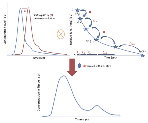
A semi-non-parametric approach to DSC perfusion analysis that offers the flexibility of a model-free (e.g., Singular Value Decomposition) estimation of the residue function, but with the regularisation that comes from using Bayesian inference applied to a parameterised function. CPI models the residue function in terms of a series of control points (subject to certain prior constraints) using spline interpolation. Perfusion and associated haemodynamic parameters can be extracted, along with an estimate of the residue function in every voxel.


