10 Feb 2021
Deep learning improves endoscopy video for diagnosis, training and treatment
Researchers from the Institute of Biomedical Engineering have been working on algorithms to better endoscopy video quality for more reliable diagnoses.
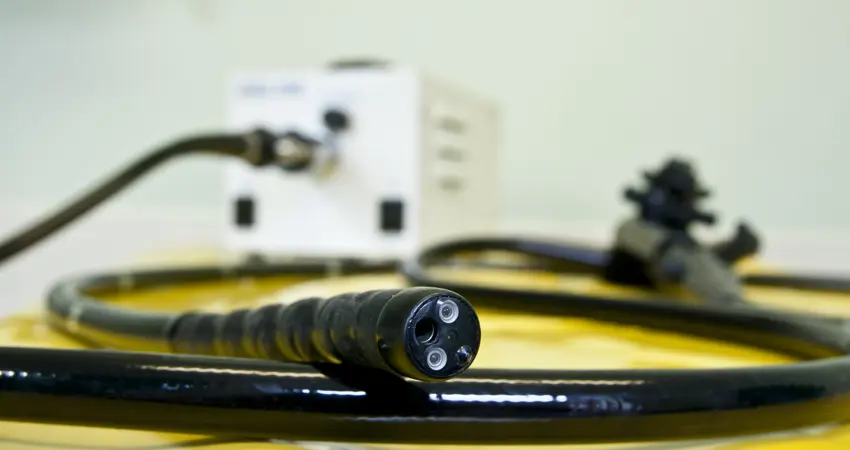
An example of an endoscope
New research from the Engineering Science Department is soon going to be used in Oxford’s John Radcliffe Hospital. The work focusses on improving the results of endoscopy, the procedure which looks at organs inside your body using a long, thin tube with a camera and light at the end, as lead author Dr Sharib Ali explains “Endoscopy is a widely used tool for diagnosis and treatment of many diseases in hollow organs.”. It can be used to examine unusual symptoms and help perform particular types of surgery, in organs such as the oesophagus, intestines and stomach.
However, there are existing problems with the method,
“Endoscopy video quality is compromised due to several factors such as tissue movement, camera motion, shaky hand of operator and interaction of light with the tissue.”
Better quality video is critical to leverage important information with higher accuracy, such as using automated methods to detect the presence or absence of lesions.
To combat these issues, Dr Ali and Prof Jens Rittscher of the Institute of Biomedical Engineering have been working on a technique that uses machine learning, “We designed a comprehensive method for identifying multiple classes of artefacts that can hinder and compromise analysis of the endoscopy video data.”. They conceptualised this idea and worked together with gastroenterologists and clinicians at the Translational Gastroenterology Unit, John Radcliffe Hospital (Prof. James East, Prof. Barbara Braden and Dr. Adam Bailey) and cancer researcher at Ludwig Institute for Cancer Research, Oxford (Prof. Xin Lu)
The quality of endoscopy video can vary widely due to the fact that the level of training of people operate the equipment. “To mitigate this problem, we also established automated assessment of endoscopy screening quality in real-time. Such a tool can be extremely beneficial for clinical training and can be used as a feedback system for endoscopists to help acquire high-quality data.”
Excitingly, the research is already going to be implemented, “The technology has been patented and soon to be used at the JR Hospital.”
Dr Ali concludes,
“The implementation and success of this research will unveil the full potential of utilising endoscopy videos for reliable analysis and improved visualisation"
"For example, the detected artefacts will be crucial to minimise false alarms of lesions. Also, the restored frames can allow more coherent visualisations of 2D or 3D mosaiced areas for intervention and therapies.”
The article is peer reviewed and published in Medical Image Analysis journal which is a leading biomedical journal with impact factor of 11.14. https://www.sciencedirect.com/science/article/pii/S1361841520302644
The work is funded by NIHR Oxford BRC.


