07 Dec 2021
Oxford researchers present new technique to probe fusion reactor armour at the nano-scale
Dr Nick Phillips and Prof Felix Hofmann, in an international collaboration, use a new technique called Bragg Ptychography to examine the nano-scale defects responsible for material degradation in future fusion reactors.

Fusion power promises an almost limitless, low carbon energy supply, which is urgently needed to address the climate emergency and meet growing energy demands. Tungsten will be used as an armour material to protect the inside of the reactor vessel from the fusion plasma. When in use, the armour components will be exposed to extreme temperatures, very high heat flux and intense ion and neutron bombardment. Unfortunately, irradiation with neutrons produced in the fusion process causes defects to form in the structure of the reactor materials. These defects can dramatically degrade the performance of armour components.
Remarkably, most of the defects irradiation produces are too small to be seen using electron microscopy, the main method used for observing material defects as small as a few atoms. While these “invisible” defects are each only a few atoms in size, high concentrations of these defects can cause dramatic changes: materials can become brittle, increase in hardness by up to 100%, reduce how well heat is transferred by more than 50% and swell, so increase in volume, by several percent.
Using X-ray beamline technology at the European Synchrotron Radiation Facility (ESRF), a team of researchers from Institut Fresnel, the Diamond Light Source, the Paul Scherrer Institute and the University of Oxford have developed a new microscopy technique that allows the measurement of these tiny imperfections at the scale of 1 billionth of a metre - the nanoscale.
“These new insights are key to developing new strategies for designing the next generation of radiation damage resistant materials”
Lead author of the study Dr. Peng Li, of Aix-Marseille University and Diamond Light Source, explains, “Our technique called Bragg Ptychography uses coherent diffraction patterns produced by a sub-micron X-ray beam rastered across the sample. From these diffraction patterns we can reconstruct the morphology of the sample with about 20 nm [nanometer] 3D resolution. Importantly, we can reconstruct in 3D how the crystal lattice within the sample has been distorted, revealing the presence of otherwise invisible defects.”
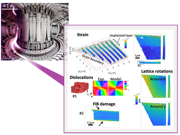
Tungsten is used to protect the inside of the reactor vessel from the fusion plasma. It is exposed to extreme heat and radiation (top left). To mimic the expected damage, Helium ion were used to bombard the a tungsten sample. The new technique, Bragg ptychography, in a micro-scale sample (right), gives new insights into the structure of defects and can be used to understand how defects are distributed in 3D. Image adapted from EUROfusion; CC BY 4.0 licence.
The key result is that, for the first time, the new reconstruction approach allows taking imperfections of the illuminating X-ray beam into account and enables researchers to see more than ever before. Prof. Virginie Chamard, of Aix-Marseille University, who has spearheaded the worldwide development of the Bragg ptychography technique, elaborates, “The new reconstruction approach is much more robust and allows crisper, higher quality reconstructions while requiring a smaller amount of experimental data. We estimate that this corresponds to an improvement in data acquisition efficiency by more than a factor of 10.”
“Our developments will be key to making this a microscopy technique that can be used by non-specialists across a wide range of instruments”
The authors applied the new microscopy technique to tungsten, the front runner material for fusion reactor armour. They were able to image in 3D the complex lattice distortions caused by helium injected into the lattice, which mimics the damage expected to form in armour components during fusion reactor operation.
Prof. Felix Hofmann from the Department of Engineering Science, University of Oxford explains, “Using Bragg ptychography has finally allowed us to actually view in 3D how atomic scale crystal defects caused by helium ion-irradiation are distributed within the tungsten matrix. Remarkably we found that fewer defects exist near grain boundaries, suggesting that perhaps nano-structured materials may offer better radiation resistance.”
“The 3D resolution of this technique has also uniquely enabled us to exclude unwanted artefacts at sample surfaces that are caused by sample preparation from our analysis. This is very important as it really allows us to pin down what defects are caused by the helium exposure. These new insights are key to developing new strategies for designing the next generation of radiation damage resistant materials.”
The future is bright when it comes to Bragg ptychography, Dr. Nick Phillips, formerly at the Oxford Department of Engineering Science, now at the Paul Scherrer Institute, concludes, “Looking ahead, our developments will be key to making this a microscopy technique that can be used by non-specialists across a wide range of instruments and we expect to improve the approach by one more order of magnitude. As more synchrotron facilities undergo upgrades, measurements like this stand to benefit enormously.”
These exciting prospects are fuelling efforts to study how atomic scale crystal defects influence the behaviour of all sorts of materials that underpin technologies central to modern life. They bring us ever closer to making commercially-viable fusion power a reality.


